31.8.06

ictotest

Thursday August 31, 2006
ICTOTEST !! (its one word)


Q; As you are reviewing the labs from patient, you noticed a note at the end of UA (urinalysis) - Ictotest positive. Do you know what is Ictotest?


A; The Ictotest® is an ultra-sensitive test to detect bilirubin in urine (detects even 0.05 mg/dL of bilirubin). It is a sensitive, reliable and easy test to diagnose bilirubinuria. Actually, it is a tablet of diazonium salt and when combines with bilirubin, it changes to blue or purple color (any other color change is a negative test). Lab run this test if there is a suspicion of bilirubinuria.

Caution: Test should be performed on fresh urine specimen and specimen should be protected from excessive light exposure. On standing, bilirubin is oxidized to biliverdin and may not react with diazonium salt tablet.

Note: Presence of Pyridium in urine may give a false positive Ictotest.

30.8.06

Wednesday August 30, 2006
FLUID FACTS

When patients return from the OR, you can get fairly good estimate of the fluid replacement in OR and assess further requirements in the ICU. Evaluate if the patient was under hydrated or over hydrated and make adjustments in fluid rates.

1. NPO Deficit:
NPO deficit = number of hours NPO x maintenance fluid requirement

2. Maintenance Fluid Requirements:
Adults: approximately 1.5 ml/kg/hr

3. Replacing Third Space Fluid Losses:

  • Superficial surgical trauma: 1-2 ml/kg/hr
  • Minimal Surgical Trauma: 3-4 ml/kg/hr (head and neck, hernia, knee surgery)
  • Moderate Surgical Trauma: 5-6 ml/kg/hr (hysterectomy, chest surgery)
  • Severe surgical trauma: 8-10 ml/kg/hr (or more) (AAA repair, nehprectomy)
4. Blood Loss:
  • Replace 3 cc of crystalloid solution per cc of blood loss (crystalloid solutions leave the intravascular space).
  • When using blood products or colloids replace blood loss volume per volume.

Example

62 y/o male, 80 kg, for hemicolectomy
•NPO after 2200, surgery at 0800
•3 hr. procedure, 500 cc blood loss
•What are his estimated intraoperative fluid requirements?

1. Fluid deficit: 1.5 ml/kg/hr x 10 hrs = 1200 ml = 1200 ml total deficit: (Replace 1/2 first hr, 1/4 2nd hr, 1/4 3rd hour).
2. Maintenance: 1.5 ml/kg/hr x 3hrs = 360 ml
3. Third Space Losses: 6 ml/kg/hr x 3 hrs =1440 ml
4. Blood Loss: 500ml x 3 = 1500ml

Total = 1200+360+1440+1500=4500ml

29.8.06

Tuesday August 29, 2006
Endotracheal Tube Cuff Pressure and VAP (Ventilator-associated pneumonia)


As we are learning more and more about dangers of Ventilator-associated pneumonia (VAP), we are in constant quest of finding factors influencing its occurance, rate and resolution.

There are 2 basic objectives for inflated cuff of ETT (endotracheal tube) 1) to abolish air leak around ETT 2) to prevent aspiration of pharyngeal contents into the trachea. ICUs use any level of cuff pressure from 20 cm H2O to 30 cm H2O in ETT ( 25 cm H2O is thought to be sufficient). The concern always is that with increasing the cuff pressure beyond any given threshold, it may compromise mucosal blood supply and may result in subglottic stenosis. Tracheal mucosal ischemia occurs when endotracheal tube cuff pressure exceeds above 34 cm H2O (some suggests it is safe till 40 cm H2O).

We learned that (and its now part of some VAP bundles) that continuous aspiration of subglottic secretions (CASS) helps in preventing VAP. Echoing on the same note Drs. Chendrasekhar and Timberlake from Trauma Services, Iowa Methodist Medical Center, Des Moines, Iowa tried to find the threshold below which subglottic secretions cross the balloon barrier. They used 10 ex-vivo tracheas obtained from previously euthanized pigs. They found that: The average ETT cuff pressure required to maintain secretions above the cuff was 29.5 ± 3.2 cm H2O 1.

This is a very small in-vivo study and in no way provide any guideline. More work is required but it tells us that this is an important aspect to explore in preventing VAP.

Automatic regulation of the cuff pressure in endotracheally-intubated patients: R. Farré and co. had previuosly reported a device that can provide automatic and continuous regulation of cuff pressure in protecting the trachea from tissue damage and for reducing the risk of ventilator-associated pneumonia
2.


Addendum: This is not a common practice in ICUs to utilize manometer to measure ETT cuff pressure. One recent study of 93 patients found that only 27% of pressures were within 20 - 30 cmH2O and actually 27% exceeded 40 cmH2O. Neither patient morphometrics, institution, experience of anesthesia provider, nor tube size influenced measured cuff pressure !! 3.

Is it time to do manometric cuff pressure everyday on our intubated patients?


References: click to get abstract/artice

1.
Endotracheal Tube Cuff Pressure Threshold for Prevention of Nosocomial Pneumonia - The Journal of Applied Research - Voulme 3 issue 3.
2.
Automatic regulation of the cuff pressure in endotracheally-intubated patients - Eur Respir J 2002; 20:1010-1013
3.
Endotracheal tube cuff pressure in three hospitals, and the volume required to produce an appropriate cuff pressure - BMC Anesthesiol. 2004; 4: 8

28.8.06

Monday August 28, 2006
Revisiting Pulmonary Artery Diastolic-Pulmonary Wedge Pressure Gradient


We don't see floatation of pulmonary artery catheter (PAC) as much as we used to see. Lets revisit one important but forgotten value obtained via PAC.

PADP - PAOP
(Pulmonary Artery Diastolic-Pulmonary Wedge Pressure Gradient).

Most of the literature in regards to this value is 15-30 years old but proven to be very easy to calculate but very vital to follow
1, 3.

Once this gradient starts to exceeds by 6 mm Hg or more, the patient has shown to have a much poorer prognosis particularly in septic patients. Probable explanation is pulmonary venous vasoconstriction induced by endotoxemia in sepsis or postcapillary lekocyte aggregation in development of ARDS
2, 4.

One study suggests that although an initial PAD-PWP gradient in patients with sepsis is associated with a high mortality, a much more sensitive indicator is to follow the trend. There was a 91% mortality in patients with persisting or increasing gradients
2.


References: click to get abstract/artice

1.
Pulmonary hypertension in sepsis: Measurement by the pulmonary arterial Diastolic-pulmonary wedge pressure gradient and the influence of passive and active factors. Chest 1978; 73:583-91

2.
Significance of the pulmonary artery diastolic-pulmonary wedge pressure gradient in sepsis. Crit Care Med 1982; 10:658-61

3.
Pulmonary artery diastolic and wedge pressure relationships in critically and injured patients. Arch Surg 1988; 123:933-6

4.
Increased Pulmonary Venous Resistance Contributes to Increased Pulmonary Artery Diastolic-Pulmonary Wedge Pressure Gradient in Acute Respiratory Distress Syndrome - Anesthesiology: Volume 102(3) March 2005 pp 574-580

27.8.06

Sunday August 27, 2006


Q; 72 year old female with no significant past medical history has been admitted from ER to ICU with progressive mental status change over last 3 days. Only significant finding in workup is Na+ of 123 mEq/liter. The only pertinent history is start of a new anti-depressant medication about 2 weeks ago. Per son, there is no sign of drug over-dose ?


A; Anti-depressants' associated hyponatremia.

One of the significant but less know side effect of anti-depressants, mostly SSRIs is hyponatremia. Exact mechansism is unknown but it causes SIADH (Syndrome of Inappropriate Antidiuretic Hormone). It has also been reported with 'atypical' anti-depressents' (venlafaxine, trazodone, maprotiline, nefazodone, bupropion) as well as with tricyclic antidepressants (TCAs) and monamine oxidase inhibitors (MAOIs).

When prescribing antidepressants particularly to elderly patients, consideration of hyponatremia should be kept in mind. Patients who develop mental status change need prompt assessment of electrolytes' status. Patients already at risk of the SIADH (such as cancer) should be prescribed anti-depressent with caution and close followup.


Related previous pearl:
ANTIDEPRESSANTS' AND SEROTONIN SYNDROME


References: click to get abstract/artice

1. Hyponatremia in the psychiatric population: a review of diagnostic and management strategies. Psychiatr Ann 2003; 33:318–325
2. Hyponatremia secondary to antidepressants. Psychiatr Ann 2003; 33:333–339
3. Hyponatremia in elderly psychiatric patients treated with selective serotonin reuptake inhibitors and venlafaxine: a retrospective controlled study in an inpatient unit. Int J Geriatr Psychiatry 2002; 17:231–237
4. Hyponatremia with venlafaxine. Ann Pharmacother 1998; 32:49–50
5.
Severe symptomatic hyponatremia during citalopram therapy - a case report - BMC Nephrology 2004, 5:2

26.8.06

Re-intubation

Saturday August 26, 2006


Q; What is the re-intubation rate in your ICU ?

A; Re-intubation rates have been reported in literature anywhere from 4% - 20% but most of the experts agree that as far as its less than 15%, you are in normal / safe zone. Ideal would be less than or equal to 5%. Other way is to keep track of reintubation rate and making sure that it is not increasing in your ICU.


Related links:

HOW TO ESTABLISH A VENTILATOR WEANING PROTOCOL , Gregory P. Marelich, MD - thoracic.org

When to wean from a ventilator: An evidence-based strategy, Ref: CLEVELAND CLINIC JOURNAL OF MEDICINE VOLUME 70, NUMBER 5 MAY 2003 page 389


Related previous pearls:

Nasal cannula vs Face mask after extubation ,

Spontaneous Breathing Trial (SBT) - how long - 30 minutes or 120 minutes?,

Bedside tip ! - Tracheal Tube Tolerance and

IV steroid to reduces postextubation stridor

25.8.06

Friday August 25, 2006
Pain management during chest tube removal !


It is important to know that chest tube removal is a painful procedure and pain could be moderate to severe in intensity. Most of the discomfort of chest tube removal relates to the initial movement of the tube.

Patient should receive some kind of analgesic prior to chest tube removal. Per one survey only 16% of nurses indicated that a prescription for pain medication was routinely available before chest tube removal
1. One study advocate administration of analgesic 20 minutes prior of removal of chest tube with either choice of morphine or ketorolac 2. Unfortunately, chest tube removal are poorly controlled with opioids and low dose morphine doesn't help much and morphine upto 4 mg may be required 2. In cases where narcotics are of concern, ketorolac is found to be a good choice. Intrapleural administration of bupivicaine did not show any benefit 3.

One interesting study was done few years ago, where EMLA (topical lidocaine 2.5% and prilocaine 2.5%) cream applied 3 hours prior to chest drain removal was more effective than IV morphine. EMLA cream can give analgesia to a depth of 5mm
4.


References: click to get article/abstract

1.
Chest tube removal practices in critical care units in the United States. - Am J Crit Care. 1995 Nov;4(6):419-24.

2.
Appropriately Timed Analgesics Control Pain Due to Chest Tube Removal - American Journal of Critical Care. 2004;13: 292-302

3.
Effects of interpleural bupivacaine on pleural chest tube removal pain: a randomized controlled trial - Am J Crit Care.1996 Mar;5(2):102-8.

4.
Topical Lidocaine - Prilocaine Cream (EMLA) for thoracostomy tube removal. Anesth Analg 1999;88:1107-8.

24.8.06

Thursday August 24, 2006
Red urine during transfusion.


Q; You have been called to evaluate a patient who developed red-urine. At bedside, you noticed pRBC transufion in progress. What would be your first few immediate responses?

A; The first thing you need to determine is whether it is a transfusion reaction (hemolysis) or a pure hematuria. Send Urine or blood for centrifuge.

The onset of red urine during or shortly after a blood transfusion may represent hemoglobinuria from acute hemolytic reaction. To distinguish it from hematuria, if freshly collected urine is centrifuged, the urine sample remains clear red. If its pure hematuria, red blood cells settle at the bottom of the tube, leaving a clear yellow urine supernatant. Similarly patient's blood with centrifuge will turn free serum as a pink color from free hemoglobin in a clotted centrifuged specimen otherwise serum will be yellow if no transfusion reaction.

Other steps to take.

1. Halt the transfusion.

2. Send donor blood and patient's blood quickly to blood bank to make sure that right blood was transfused (repeat crossmatch and type) and for antibody screen, and direct and indirect Coombs tests.

If transfusion reaction is highly suspected:

3. Administer IV Benadryl, IV steroid, IV saline followed with IV lasix or with low dose dopamine to improve renal blood flow. Symptomatic treatment with acetaminophen.

4. Airway protection and if seems to be anaphylactic reaction, administer epinephrine (nebulizer treatment, SQ or IV drip depending on severity). Oxygen to keep saturation up.

5. Send complete lab workup including lytes, renal function (BUN/Cr), serum bilirubin level (peaks in 3-6 hours), Haptoglobin (binds to hemoglobin) , urine for hemoglobinuria, a repeat CBC (fails to show the rise in hematocrit because of intravascular or extravascular hemolysis) and DIC panel.

6. Hematology consult.
Management is largely supportive.

Related previous pearl:
TRALI
23.8.06

Wednesday August 23, 2006
Is Dopamine phasing out?


Standard guidelines regarding vasopressor and inotropic support in septic shock states: "Either norepinephrine or dopamine (through a central catheter as soon as possible) is the first-choice vasopressor agent to correct hypotension in septic shock." 1 But overall trend is towards using norepinephrine as the first-choice vasopressor to correct hypotension in septic shock after fluid resuscitation 2, 3, 4.

In March issue of Critical Care Medicine, part of SOAP (Sepsis Occurrence in Acutely Ill Patients) study looked into multiple-center observation of 1,058 patients with shock
5.

Patients were followed up until death, until hospital discharge, or for 60 days. Of patients in shock, 375 received dopamine (dopamine group) and 683 never received dopamine. The dopamine group had higher ICU (42.9% vs. 35.7%,) and hospital (49.9% vs. 41.7%,) mortality rates. It also showed diminished 30 day-survival in the dopamine group. In a multivariate analysis, dopamine administration was found to be an independent risk factors for ICU mortality in patients with shock.

Study suggests that dopamine administration may be associated with increased mortality rates in shock.


Related previous pearls:

Norepinephrine or Dopamine ? ,
Dopamine-S and Dopamine-R patients ? ,
Renal dose Fenoldopam ? and
Renal Dose Norepinephrine !


References: click to get abstract/article

1.
Vasopressor and inotropic support in septic shock: An evidence-based review Critical Care Medicine: Volume 32(11) Supplement November 2004 pp S455-S465
2.
THE SAFETY OF DOPAMINE VERSUS NOREPINEPHRINE AS VASOPRESSOR THERAPY IN SEPTIC SHOCK - chest, 2005
3.
Renal Dose Norepinephrine! - Chest. 2004;126:335-337
4.
Renal Effects of Norepinephrine in Septic and Nonseptic Patients - Chest. 2004;126:534-539.
5.
Does dopamine administration in shock influence outcome? Results of the Sepsis Occurrence in Acutely Ill Patients (SOAP) Study - Critical Care Medicine. 34(3):589-597, March 2006

22.8.06

Tuesday August 22, 2006
Xifaxan (Rifaximin)

Rifaximin is in use in other countries since last 2 decades but has been approved by FDA in USA just couple of years ago. Rifaximin was invented initially in Italy for the treatment of travelers' diarrhea but quickly found its way for improvement in hepatic encephalopathy instead or beside lactulose and neomycin 1, 2,5,6,7. Some new studies showed that it may be more effective than lactulose 3 or neomycin 4.

Atleast one multi-center trial is underway
8, as well as other trials looking at Rifaximin's efficacy in Irritable Bowel Syndrome, Clostridium Difficile-Associated Diarrhea and Ulcerative Colitis 8.

Rifaximin is an oral, semi-synthetic, nonsystemic antibiotic. The recommended dosage in hepatic encephalopathy is 1200 mg a day in divided doses.


References: click to get abstract/article

1.
Rifaximin in the treatment of chronic hepatic encephalopathy - Curr Med Res Opin. 1995;13(5):274-81.
2.
Rifaximin, a non-absorbable rifamycin, for the treatment of hepatic encephalopathy. A double-blind, randomised trial. - Curr Med Res Opin. 1997;13(10):593-601.
3.
Comparison of rifaximin and lactitol in the treatment of acute hepatic encephalopathy: results of a randomized, double-blind, double-dummy, controlled clinical trial - J Hepatol. 2003 Jan;38(1):51-8
4.
Neomycin should not be used to treat hepatic encephalopathy - BMJ 2001;323:233 ( 28 July )
5. Management of Hepatic Encephalopathy: Role of Rifaximin - Chemotherapy 2005;51 (Suppl. 1):90-95
6. Evaluation of the efficacy and safety of rifaximin in the treatment of hepatic encephalopathy: a double-blind, randomized, dose-finding multi-centre study. Eur J Gastroenterol Hepatol 2000; 12: 203-208
7.
Rifaximin, a nonabsorbed oral antibiotic, in the treatment of hepatic encephalopathy: antimicrobial activity, efficacy and safety. Reviews in Gastroenterological Disorders, 5 (Supplement 1). S10-S18.
8.
Rifaximin trials - clinicaltrials.gov

21.8.06

THAM

Monday August 21, 2006
THAM


Introduction: In patients with acute respiratory distress syndrome (ARDS), permissive hypercapnia is a strategy to decrease airway pressures to prevent ventilator-induced lung damage by lowering tidal volumes and tolerating higher arterial carbon dioxide tension. A pure respiratory acidosis generally does not require alkali therapy. Alkali therapy is indicated for either a metabolic acidosis or a mixed acidosis. The choice of buffer is based on the type of acidosis, cardiorespiratory status, and lung mechanics.

Problem with NaHCO3: Slow infusions of NaHCO3 can be used to treat non-anion gap metabolic acidosis and some forms of increased anion gap acidosis. But using NaHCO3 to treat type A (hypoxia-related) lactic acidosis can be hazardous, particularly under conditions of hypoxemia, inadequate circulation, and limited alveolar ventilation.

THAM: Under above circumstances, THAM is the preferable buffer because it does not increase PaCO2 and is excreted by the kidneys. Tromethamine (THAM) is a sodium-free alkalinizing agent that acts as a hydrogen ion (proton) acceptor. It is a weak base that combines with hydrogen ions from carbonic acid to form bicarbonate and cationic buffer. Administration of tromethamine decreases hydrogen ion concentration, which results in a decrease in carbon dioxide concentrations and an increase in bicarbonate concentrations. The administration of Tham also increases urine output through osmotic diuresis. Excretion of electrolytes and CO2 is also increased. Urine pH is raised along with the excretion of electrolytes.

Usual Dose:
Dose in ml's of 0.3M THAM = (1.1) (Wt. in Kg) (normal HCO3 – Pt’s HCO3)
OR
Dose in ml’s of 0.3M THAM = body wt in kg X base deficit in MEq/L x 1.1

Total dose should be administered over a period not less than 1 hour via central line. .3M THAM solution is available as premix and is contra-indicated in renal failure, anuria and hyperkalemia. It may cause transient hypoglycemia and respiratory depression.

20.8.06

Heated Humidifier

Sunday August 20, 2006
Does heated humidifier reduces the risk of ventilator associated pneumonia (VAP) ?


Most of the VAP bundle guidelines does not include the type of humidifier to reduce the risk of VAP, although thats the first therapy in direct contact with respiratory tract.

In latest issue of Critical Care (
ccforum.com) - The fully formatted PDF version not published yet but early version can be seen here - a study of 104 patients, who required mechanical ventilation more than 5 days, has been published. Patients were assigned either to heat and moisture exchangers (HME) (n = 53) or heated humidifiers (HH) (n = 51) * 1.

VAP was found in 8 of 51 (15.69 %) patients in the HH group and in 21 of 53 (39.62 %) in the HME group. The median time free of VAP were 20 days for HH and 42 days for HME group.

Study concluded that: The patients mechanically ventilated during more than 5 days developed a lower incidence of VAP with a heated humidifier than heat and moisture exchanger.


* HH: In the active humidifiers, called heated humidifiers (HH): the inspired gas passes across or over a heated water bath. In this study HH was provided at 37º C and 100 % relative humidity to the proximal airway (containing approximately 44 mg of water/L of gas).


HME: Passive humidifiers, called heat and moisture exchanger (HME), trap heat and humidity from the patient’s exhaled gas and returns some of that to the patient on the subsequent inhalation.


Related previous pearls:
Elevation of the head of the bed- 30 or 45 degrees ?
Intrahospital transport - a risk factor for VAP ? and
Oral care in ICU


Helpful links:
IHI's Implement the Ventilator Bundle and
7 strategies to prevent VAP: a look at the evidence (source: todayshospitalist.com)


Reference: click to get abstract

Ventilator-associated pneumonia using a heated humidifier or a heat and moisture exchanger - a randomized controlled trial - Critical Care 2006, 10:R116

19.8.06

Pulmonary artery perforation

Saturday August 19, 2006


Q: You have been called by the nurse at bedside as she noticed some fresh blood in ETT. Patient has pulmonary artery catheter (PAC) in place. You are worried about pulmonary artery perforation. What would be be your first response before you initiate whole 9 yards of workup and management?


A; "Get good lung up".

First thing you need to know which pulmonary artery (Right or Left) has distal end of PAC and get patient 's position change to lateral decub. with good lung up to avoid soiling of good lung.

If patient is on ventilator, other trick is to increase PEEP (literature is controversial about its efficacy but there is no harm in increasing PEEP to control catastrophy). Overall management is supportive if pulmonary artery perforation is confirmed. Actually it is better to leave PAC intact with ballon in inflated position !! Reversal of anticoagulation if needed, hemodynamic support and preventing good lung from soiling (like applying double lumen ETT) are the initial mainstay of treatment - followed by attempt to control bleeding with interventional radiology help.


Read article:
Pulmonary Artery Rupture Induced by a Pulmonary Artery Catheter: A Case Report and Review of the Literature. Alexandre R. Abreu, MD, Michael A. Campos, MD and Bruce P. Krieger, MD Division of Pulmonary and Critical Care Medicine, Department of Medicine, University of Miami School of Medicine, Miami, Florida. Ref: Journal of Intensive Care Medicine, Vol. 19, No. 5, 291-296 (2004)

18.8.06

Airtraq


Friday August 18, 2006
AIRTRAQ !

We try to keep intensivists abreast with new technologies. Orotracheal intubation is one of the most essential but could be the most frustrating procedure in ICU.

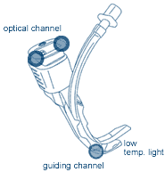
Airtraq is an intubation device which provides a magnified angular view of the larynx and adjacent structures. No hyperextension of the neck is required (good with spine surgery) and actually you can do intubation in any position. It prevents dental trauma and avoid multiple intubation attempts with quick learning curve. It is a great tool for very obese patient or presumed difficult intubation.

Airtraq has two separate channels:

The optical channel: contains a high definition optical system.
The guiding channel: holds the endotracheal tube (ETT) and guides it through the vocal cords.


It can be used with any standard endotracheal tube.

Visit website for video and related articles at
www.airtraq.com


Note: icuroom.net has no financial relationship with any company. Info provided here is 100% for educational purpose.

17.8.06

TRALI

Thursday August 17, 2006
TRALI

Transfusion-Related Acute Lung Injury (TRALI) - a different condition from transfusion related pulmonary edema - has an incidence of approximately 1:5,000 units of blood transfused. Approximately 6% of the patients die from TRALI and 72% require mechanical ventilation. It typically occurs within 2 – 4 hours after infusion of blood product, although it can occur up to 40 hours following the transfusion. Most cases resolve spontaneously within 3 – 4 days and there are rarely long time sequelae.

The etiology of TRALI is incompletely understood. TRALI can occur after transfusion of packed RBCs, whole blood, platelets, fresh frozen plasma and cryoprecipitate.

Treatment is supportive, although high-dose steroids have been shown to be successful in old animal studies. In patients with documented TRALI, ideally, subsequent transfusions should be autologous, or in case of packed RBCs, washed to remove residual plasma.


Read review article:
Transfusion-Related Acute Lung Injury - Ref: Mayo Clin Proc. 2005;80:766-770

16.8.06

A-line and volume

Wednesday August 16, 2006
Bedside Tip - Need volume?


Even Before you start monitoring CVP (central venous pressure) or waiting for urine to start trickle in bag with IVF bolus, just a glance at ARTERIAL WAVEFORM may tell you that your patient is volume depleted. The arterial trace may reveal the intravascular fluid depletion by an exaggeration of the decrease in pressure (pulsus paradoxus) that normally occurs during or shortly after inspiration. A difference of 10 mm Hg or higher from inspiration to expiration identifies pulsus paradoxus. It may even occur before parameters such as central venous pressure, blood pressure, or urine output substantially decrease, in both ventilated or spontaneously breathing patient
1.

Interesting question raised in one of the article related to this topic
1 : Whether a goal-directed therapy taking into account the assessment of the respiratory variation in arterial pressure may improve the outcome of mechanically ventilated patients with shock.....?. Click reference # 1 to see waveforms and algorithm.


Related:
We found 2 good review articles on basic understanding of arterial waveform to distribute among residents and new fellows (its august - right?)

1.
HEMODYNAMIC MONITORING: DYNAMIC RESPONSE ARTIFACTS - Michael L. Cheatham, MD, FACS, FCCM - Director, Surgical Intensive Care Units, Orlando Regional Medical Center, Orlando, Florida.

2.
Monitoring Arterial Blood Pressure: What You May Not Know - Ref: Critical Care Nurse. 2002;22: 60-79


Reference: click to get full article

1.
Changes in Arterial Pressure during Mechanical Ventilation. Anesthesiology. 103(2):419-428, August 2005. Michard, Frederic M.D., Ph.D.

15.8.06

Tuesday August 15, 2006


Q: What volume of chest tube drainage will make you comfortable to consider removing it?


A:
Once your drainage comes down to less than or equal to 200 ml over 24 hours without any evidence of air leaks - you may be in safe zone to discontinue chest tube
1.


Read excellent review on
chest tube management (pdf file) from www.surgicalcriticalcare.net (Department of Surgical Education, Orlando Regional Medical Center).


Reference: click to get abstract

1.
When to remove a chest tube?: A randomized study with subsequent prospective consecutive validation - J Am Coll Surg. 2002; 195:658-662.

13.8.06

Monday August 14, 2006
Temporal Artery Infrared Thermometry !

As we all know that there are many different ways to measure temperature but what is the most accurate way particularly in ICU ?.

"Temporal Artery Infrared Thermometry" (
TemporalScanner - Exergen ) appears to be more accurate than ear or rectal thermometer in responding to change in fever. It is non-invasive, gentle, fast, easy and reliable to use.

From ICU perspective, there are 3 important points:

  • It is as accurate as a pulmonary artery catheter 1.
  • It responds faster than temperature taken rectally 2.
  • New version contain anti-microbial head to prevent transmission of VRE and MRSA.


Also, it is said to be 90% more cost-effective.


It captures the naturally emitted heat from the skin over the temporal artery, taking 1,000 readings per second, selecting the most accurate. Automatically its Arterial Heat Balance system adjusts the small temperature loss from cooling at the skin.

You can watch the video
here (mpeg).


Note: icuroom.net has no financial relationship with any company. Information provided here is 100% for educational purpose.


Refereces:
1. A Comparison of Measurements from a Temporal Artery Thermometer and a Pulmonary Artery Catheter Thermistor. National Conference of Clinical Nurse Specialists, Poster Section, Atlanta, GA March 14-16, 2002. - Carroll DL, Finn C, Gill S, Sawyer J, Judge B (Massachusetts General Hospital).


2. When body temperature changes, does rectal temperature lag behind? PAS Annual Meeting, Baltimore MD, May 4-7, 2002 - Greenes DS, Fleisher GR. Division of Emergency Medicine, Children's Hospital, Harvard Medical School, Boston, MA.

Sunday August 13, 2006
Antibiotics and our failure !

About a week ago, we posted a remarkable study done by Dr. Anand Kumar and coll. and recently published in Critical Care Medicine * which found that
"Administration of an antimicrobial within the first hour of documented hypotension was associated with a survival rate of 80% and time to initiation of effective antimicrobial therapy was the single strongest predictor of outcome ." Unfortunately, same study found that "only 50% of septic shock patients received effective antimicrobial therapy within 6 hrs of documented hypotension !"

*
Get antibiotics quickly !! - our previous pearl


On the same theme, a study published in July 2006 issue of chest
1 looked into the factors shielding physicians from administrating antibiotics early in community-acquired pneumonia (CAP). Study found that the 4 major factors which mislead physicians are:

  1. Altered mental state,
  2. Absence of fever,
  3. Absence of hypoxia and
  4. Increasing age

Another major significance of this study - Editors' note: This is probably the first study which challenges the notion adopted by The Joint Commission of Health Care Organizations (JCAHO) and the National Quality Forum that delay of antibiotics administration was directly associated with an increased mortality. Rather, study argues that prolonged TFAD (time to the first antibiotic dose) is more a marker for comorbidities due to the fact an altered mental state and the absence of fever were stronger predictors of mortality than a TFAD of more than 4 hours.
Study questions: It is very appropriate to administer antibiotics as soon as possible but why a difference of a few hours in administration will impact adversely on mortality particularly as most patients have had symptomatic disease for several days?


Few important observations in the study

* TFAD is likely to be a marker of comorbidities driving both an atypical presentation and mortality rather than directly contributing to outcome.

* In elderly patients, and especially those presenting with confusion, physicians need to be more vigilant in excluding a diagnosis of pneumonia.

* An altered mental state may not only make it difficult for physicians to make a diagnosis, it may also delay presentation to hospital.


Referece: Click to get abstract
1.
Delayed Administration of Antibiotics and Atypical Presentation in Community-Acquired Pneumonia - Chest. 2006;130:11-15.

12.8.06

Saturday August 12, 2006
Drug bug - 2

Yesterday we learned that ratio solution is always between gram and millilitres (gms:mls - 1: 1000).

Now let see: What does percentage (%) expresses ?

Percentage (%) expresses the number of grams of the drug per 100 milliliters of solution.


Examples:

· Calcium Gluconate 10% contains 10 gms 'pure drug' per 100 mL solution

means
10 gm / 100 mL or
10,000 mg / 100 mL or
100 mg / mL


· Lidocaine 2% contains 2 gm 'pure drug' per 100 mL

means
2 gm / 100 mL
2000 mg / 100 mL
20 mg / mL

Notice that percentage is written between 2 unequivalent measurements (gram and millilitre) - drug bug ?

11.8.06

Friday August 11, 2006
Drug bug !

Intensivists should have full command of any drug, its concentration and its infusion protocols, that is ordered in ICU. If some rules are followed, there is little chance that you will make a mistake. For example

Do you know the standard ratio solution?

Ratio solution express the number of grams of the drug per total millilitres of solution.
(Note: This is NOT mg to ml but gram to mls - got it?)

Example:

· Epinephrine 1:1000 contains 1 gram 'pure drug' per 1000 mL of solution.

means
1 gm : 1000 mL or
1000 mg : 1000 mL or
1 mg : 1 mL
- but always written as gram : millilitres


- drug bug?

Thursday August 10, 2006
Level of central line tip may predispose to thrombosis

Interesting retrospective review of 428 central lines (inserted into 334 patients) was done in UK to look into the level central line tip's relation to thrombosis 1. The median follow-up was 72 days.

* The chest radiograph obtained post-catheter insertion, as well as follow-up radiographs, linograms, venograms and Doppler ultrasounds (US), were reviewed.

They found that: "There was a significant difference in thrombosis rate between lines sited with the tip in a distal third of the superior vena cava (2.6%) compared with a proximal third of the superior vena cava (41.7%) - CVC with tips in a proximal position were 16 times more likely to thrombose than those in a distal position".


Related previous pearls:

  1. CXR reading for optimum tip of central line
  2. What if SC central line ends up in IJ vein?
  3. Peres-nomogram

Reference:
1. To clot or not to clot? That is the question in central venous catheters - Clinical Radiology, Volume 59, Issue 4, April 2004, Pages 349-355

9.8.06

Wednesday August 9, 2006
External Jugular (EJ) approach to central venous line

Being an intensivist, it is important to know the pros and cons of EJ central venous catheterization.


Disadvantages

  • EJ vein is not in a direct line with the right atrium.
  • a valve is frequently present at the junction of the EJV and the SC vein.
  • It has a circuitous course and less commonly it enters directly either in the subclavian vein or internal jugular vein.
  • 10% of the time its hard to feel free flow of wire and to thread J-wires past the clavicle and also due to presence of valves in vessel 2.
  • Hydrothorax has been described in literature with left EJ approach 1.

Due to above facts

  • It is not a prefered approach.
  • It is not a reliable way to do CVP (central venous pressure) monitoring.
  • It is not a good approach if you decide to float pulmonary artery catheter later.

Advantages

* It is an approach for use in a heavily anticoagulated or thrombocytopenic patients because it is easy to apply pressure to the site of venepuncture - afterall the external jugular vein is a peripheral vein.

* Recently, a small study of 23 patients described the 'right' EJV as an acceptable and prefered access site when the right IJV is not available for central line due to various reasons such as existing Hickman catheter in the right IJV or nearby tracheostomy tube
3.


Bonus Pearl:There is an inverse correlation between the size of the external and internal jugular veins, so if you see a large EJV on patient's neck, expect potentially more difficult internal jugular venepuncture 4.


References: click to get abstract/article

1.
Bilateral hydrothorax caused by left external jugular venous catheter perforation - J Clin Anesth. 1994 May-Jun;6(3):243-6
2.
Shoulder manipulation to facilitate central vein catheterization from the external jugular vein - Anaesth Intensive Care.1991 Nov;19(4):567-8
3.
Use of the Right External Jugular Vein as the Preferred Access Site When the Right Internal Jugular Vein Is Not Usable - Journal of Vascular and Interventional Radiology 17:823-829 (2006)
4.
Prediction of a small internal jugular vein by external jugular vein diameter
- Anaesthesia 1997 Mar;52(3):220-2

8.8.06

Tuesday August 8, 2006


Q; After successful completion of Transjugular Intrahepatic Porto-systemic Shunt (TIPS) for variceal bleeding - hepatic encephalopathy __________ ?

A) tends to get better

B) tends to get worse

C) It has nothing to do with TIPS


Answer is B:

Hepatic encephalopathy tends to get worse after successful completion of TIPS as due to shunting, blood flow to the liver is reduced, which might result in increase toxic substances reaching the brain without being metabolized first by the liver. It can be treated medically such as diet, lactulose or by narrowing of the shunt by insertion of a reducing stent.


References: click to get abstract/article

1.
Treatment for hepatic encephalopathy: tips from TIPS? - Journal of Hepatology 42 (2005) 626–628

2.
Hepatic encephalopathy after TIPS-- retrospective study - Vnitr Lek. 2002 May;48(5):390-5
3.
TIPS for Prevention of Recurrent Bleeding in Patients with Cirrhosis: Meta-analysis of Randomized Clinical Trials - Radiology. 1999;212:411-421

7.8.06

Monday August 7, 2006
rhAPC (Xigris) and low-dose heparin in septic shock


Interesting laboratory work from New York, published this month in Critical Care Medicine issue 1, regarding possible interaction of rhAPC (recombinant human activated protein C) and low dose heparin. Following preparation was made in 3 steps:

  1. Neutrophils and platelets are taken from normal (control) subjects,
  2. Stimulated with plasma and serum from 21 patients in septic shock and
  3. Perfused over endothelial cells.

Now, 3 groups were made with addition of each of the following, suspended in septic plasma.

  • Activated protein C, or
  • low-dose heparin, or
  • low-dose heparin with rhAPC

What they found:

Activated protein C significantly decreased neutrophil adhesion and aggregation and increased rolling velocity in cells stimulated with both septic serum and septic plasma. Significant decreases in platelet-neutrophil aggregates induced by septic plasma were also observed.

Low-dose heparin alone had no effects on these variables.

But

The addition of low-dose heparin to cells suspended in septic plasma and rhAPC attenuated the benefits observed with rhAPC alone in each of these variables.

This is only one work but raises the question - Should we hold on using the low dose heparin as DVT prophylaxis with rhAPC {Drotrecogin alfa (activated) - Xigris} ?


* Low-dose heparin is used as unfractionated heparin in a concentration of 0.15 IU/mL. This heparin concentration, represents the concentration used for deep vein thrombosis prophylaxis, was derived as a fraction of the concentration, 0.2-0.7 IU/mL, used for full anticoagulation.


Reference: click to get abstract/article

Effect of recombinant activated protein C and low-dose heparin on neutrophil-endothelial cell interactions in septic shock - Critical Care Medicine. 34(8):2207-2212, August 2006.

5.8.06

Saturday August 5, 2006

Q; Where would you measure the PAOP (pulmonary artery occlusion pressure) ?

A: Remember !. The pulmonary artery occlusion (`wedge') pressure should be read at the end-expiratory point. This seems easy but at bedside its get tricky and often forgeted as it may be different in spontaneously breathing and patient on positive pressure ventilation.

If patient is breathing spontaneously, this pressure will correspond to the 'peak' of the wedge pressure trace (first strip),

If patient is on positive pressure ventilation, it will correspond to the 'trough' (second strip).

(Just remember - ventilator put people down)


First strip is - spontaneously breathing patient
Second strip is - patient on positive pressure ventilator

4.8.06

Friday August 4, 2006
Get antibiotics quickly !!

A remarkable work of review of medical records of 2,731 patients with septic shock from Dr. Anand Kumar and coll. is recently published in Critical Care Medicine 1. The main outcome measure was survival to hospital discharge.

What they found - "Administration of an antimicrobial effective for isolated or suspected pathogens within the first hour of documented hypotension * was associated with a survival rate of 80%".

* Hypotension was defined as a mean blood pressure of less than 65 mm Hg, a systolic blood pressure of less than 90 mm Hg, or a decrease in systolic pressure of 40 mm Hg from the patient's baseline.

  • Each hour of delay in antimicrobial administration over the ensuing 6 hrs was associated with an average decrease in survival of 7.6%.
  • In multivariate analysis, time to initiation of effective antimicrobial therapy was the single strongest predictor of outcome.


But look how good we are performing in field. Data found that only 50% of septic shock patients received effective antimicrobial therapy within 6 hrs of documented hypotension !!!


Reference: click to get abstract

Duration of hypotension before initiation of effective antimicrobial therapy is the critical determinant of survival in human septic shock - Critical Care Medicine. 34(6):1589-1596, June 2006

3.8.06

Thursday August 3, 2006
3 Cs of Intensivist !


Q; What makes a 'successful' Intensivist ?

A; 3 Cs

  • Communicative
  • Co-operative
  • Cost-effective

Wednesday August 2, 2006
In hypertensive intracerebral hemorrhage (ICH) !

Stroke Council of the American Heart Association recommends that goal of mean arterial pressure (MAP) in patients with ICH is at less than 130 mm Hg, and if ICP monitor is in place, to maintain cerebral perfusion pressure (CPP) of more than 70 mm Hg, to reduce hematoma expansion 1.

- CVP should be maintained between 5-12 mm Hg or pulmonary wedge pressure at 10-14 mm Hg.
- If systolic arterial blood pressure falls below 90 mm Hg, pressors should be given.


Last year Dr. Adnan Qureshi and coll. from UMDNJ - University of Medicine and Dentistry of New Jersey, Newark, NJ, reported that patients treated within 6 hours * of symptom onset were more likely to be functionally independent at 1 month compared with patients who were treated between 6 and 24 hours 2. On followup to this, last month, Dr. Qureshi and coll. reported study of 46 patients (29 patients were treated), - with intravenous nicardipine 3.The onset of intravenous nicardipine is within 1 to 5 mins and the effect last from 10 to 15 mins.

  • Intravenous nicardipine was initiated at a rate of 5 mg/hr.
  • If the MAP was not reduced to less than 130 mm Hg after 15 mins, the infusion dose was increased by 2.5 mg/hr.
  • The 2.5-mg/hr increments continued every 15 mins until the maximum dose of 15 mg/hr was reached.
  • Once the target blood pressure was reached, the infusion rate was decreased to 3 mg/hr and subsequently adjusted by 1 to 2.5 mg/hr to maintain the MAP in the targeted range (between 100 and 130 mm Hg).
  • The patients with previous meds that have hypotensive properties were continued on the preadmission dose such as beta-blockers.
  • Intravenous nicardipine was discontinued in the event of serious adverse event.


They looked into tolerability, neurologic deterioration, hematoma expansion and 30-days functional outcome. Group found that MAP goals recommended by the American Heart Association using intravenous nicardipine are well tolerated in patients with ICH. Also, they did not find any evidence to suggest that patients with ICH who received intravenous nicardipine within 24 hrs of symptom onset had excessively high rates of neurologic deterioration or hematoma expansion.


* A multiple-center clinical trial, Antihypertensive Treatment of Acute Cerebral Hemorrhage is underway to study this approach within 6 hrs of symptom onset 4.


References: click to get abstract/article

1.
Guidelines for the management of spontaneous intracerebral hemorrhage: A statement for healthcare professionals from a special writing group of the Stroke Council, American Heart Association. Stroke 1999; 30:905-915

2.
A Prospective Multicenter Study to Evaluate the Feasibility and Safety of Aggressive Antihypertensive Treatment in Patients With Acute Intracerebral Hemorrhage - Journal of Intensive Care Medicine, Vol. 20, No. 1, 34-42 (2005)

3.
Treatment of acute hypertension in patients with intracerebral hemorrhage using American Heart Association guidelines - Critical Care Medicine. 34(7):1975-1980, July 2006.

4.
Antihypertensive Treatment in Acute Cerebral Hemorrhage trial

1.8.06

Tuesday August 1, 2006


Q; Which order you should add while writing orders for pRBC transfusion beside rate ?


Answer; "Please insert 18 gauge IV catheter if possible for pRBC transfusion".

In adult patient if available, pRBC should be transfused preferably through atleast 18 gauge catheter. This is not a guideline or a necessity but an 18-G catheter provides good flow rates for cellular components.

On the flip side, drawing blood from smaller catheter can produce hemolysis and lead to over-diagnosis of hyperkalemia and possible patient safety issue. As the catheter gets smaller, hemolysis goes up 1.

24 gauge (100% chance of hemolysis),
22 gauge (25% chance of hemolysis),
20 gauge (15% chance of hemolysis),
18 gauge (10% chance of hemolysis),
14 -16 gauge (0% chances of hemolysis).


Related: Do you have a blood transfusion protocol in your ICU?.

See this
Blood Transfusion Protocol from stanford, designed to minimize the over and unnecessary pRBC transfusions.


Reference:

1.
A comparison of hemolysis rates using intravenous catheters versus venipuncture tubes for obtaining blood samples - J Emerg Nurs.1996 Dec;22(6):566-9